Works

Kidney stone (urolithiasis)
Kidney stoneswere traditionally analyzed after being ground into powder for chemical analysis. Thin-section preparation now makes it possible to analyze internal structures by tissue type and to observe growth patterns within the stone.
( The image below is a Winner of the Special Arts Award, 2023 NIKON JOICO AWARD_on Osaka Univ. Dr. Maruyama's article)

Carbon fiber reinforced plastic (CFRP prepreg)
CFRP is a lightweight, high-strength material used in many applications, including aircraft and golf shafts. With thin-section preparation, internal features that are difficult to observe in conventional sections—such as voids and fiber/resin distribution—can be seen clearly.


Lasiotrichius succinctus chest
Lasiotrichius succinctus (pronotum / thorax)
Scale: 1 mm


Corymbia succedanea abdomen
With thin-section preparation, even the jelly-like internal tissues of insects—once considered difficult to observe—can be examined clearly. The image below shows a magnified detail.


Carcharhinus galapagensis
Shark skin is sometimes used like a grater, and this thin section shows why: each dermal denticle rises as a hook-like structure on the surface, and blood vessels can be seen running within.

Trypoxylus dichotomus horn
Insect cross-sections that were difficult to observe with conventional biological sectioning can be examined using our resin-embedded thin-section method. Overlapping layers of the beetle cuticle—previously hard to distinguish—can now be clearly seen and even counted. Scale: 1 mm

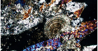
Ophiopholis mirabilis
Thin-section preparation enables observation of an animal’s hard and soft tissues on the same plane. The colors are natural—no staining is used.

around the mouth of Ophiopholis mirabilis
The radula and the surrounding muscles can be clearly observed.

a tubercular process on the arm of Ophiopholis mirabilis
It is a tubercle-like hard tissue on the surface of the arm. The way the skeletal elements connect highlights the beauty of natural form.

attachment part of muscles and spicules of Ophiopholis mirabilis
You can observe the attachment area between the arm ossicles and the muscles.

bowel of Ophiopholis mirabilis
The brown area is the stomach. The folded (plicated) structure of the stomach wall is clearly visible.